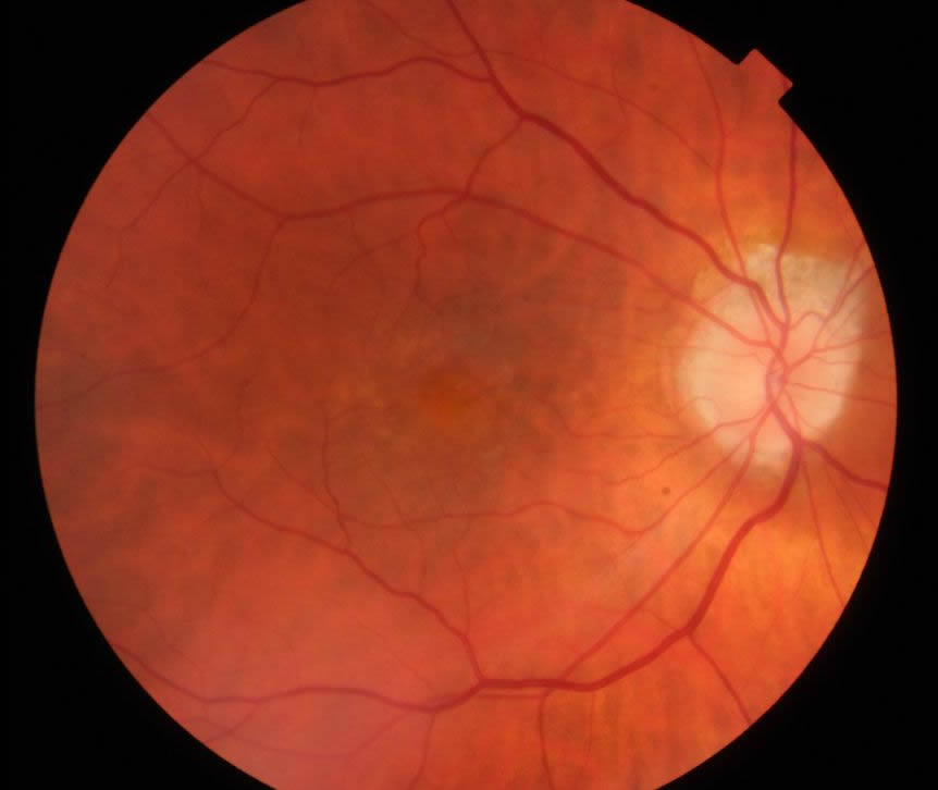
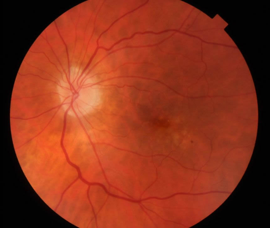
Fundus photography is a non-invasive diagnostic imaging technique used to document the retina; the neurosensory tissue of the eye that transmits the optical images we see into the electrical images our brain understands.
These photographs give one of the best interpretations of overall retinal health. In a fundus photograph, the retina, the central macula, and optic nerve are visualized. This image assists the ophthalmologist in the diagnosis, documentation, and treatment of retinal disorders.
What can I expect during a fundus photograph?
While the test is being performed, you will be positioned in front of the camera. You may see some after effect colors from the filters used while imaging your eyes, but they will disappear within minutes just like a normal family portrait flash. No possible adverse reactions will occur.


