Vascular endothelial growth factor, or VEGF, is a molecule generated by the body that causes new blood vessels to grow. Sometimes this is normal; however, in retinal disorders such as macular degeneration or macular edema from diabetic retinopathy or vein occlusions, the eye produces too much VEGF. This higher level of VEGF causes abnormal blood vessels to grow and causes them to leak fluid or to bleed, causing distortion of vision. Injections of anti-VEGF drugs into the eye block the activity of VEGF and often result in a decrease in the fluid or bleeding caused by the abnormal vessels. This procedure only takes moments and is done in the office.
What Anti-VEGF Drugs are available?
Anti-VEGF therapy options are based upon individual retinal disorders in combination with diagnostic test results.
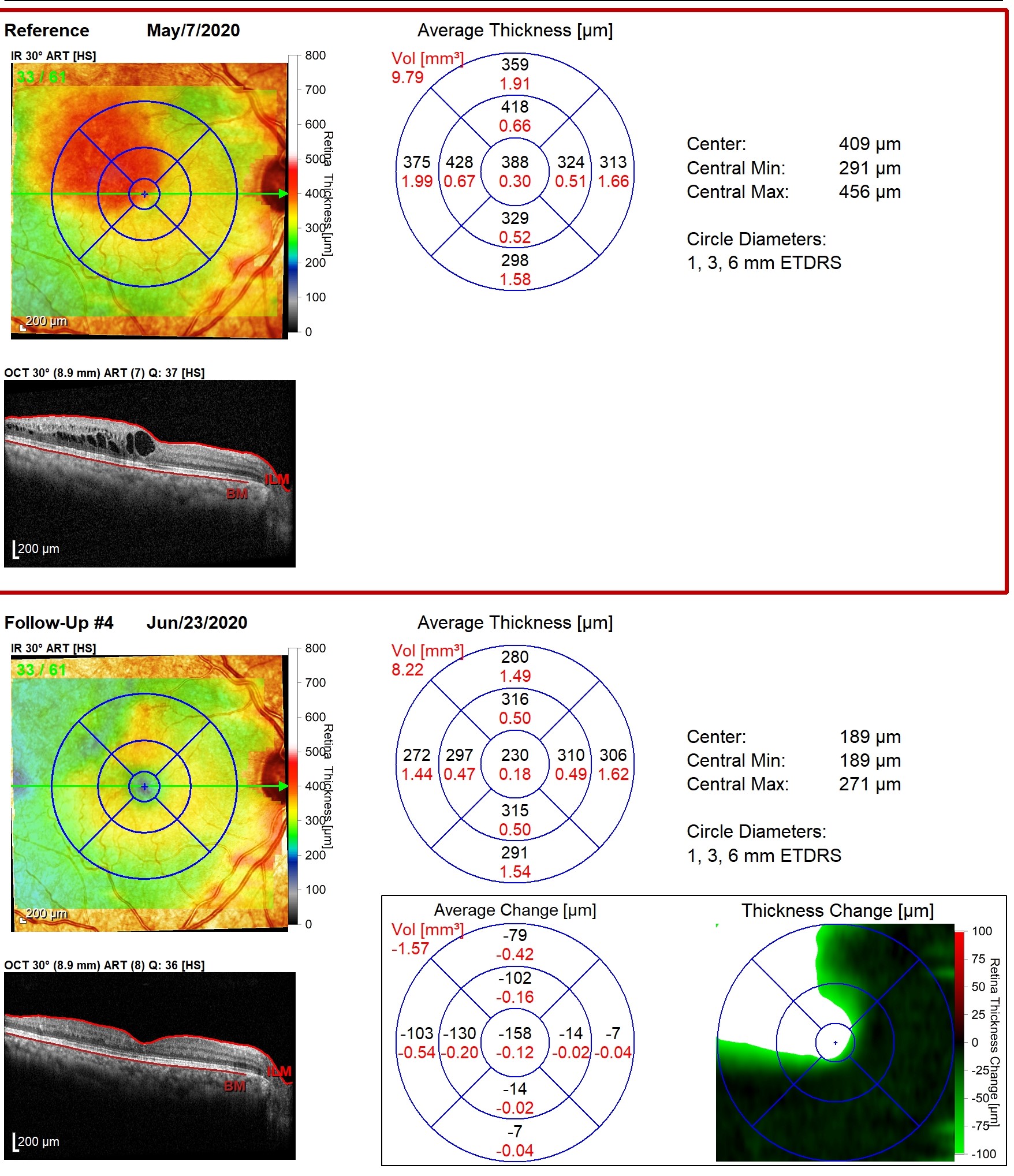
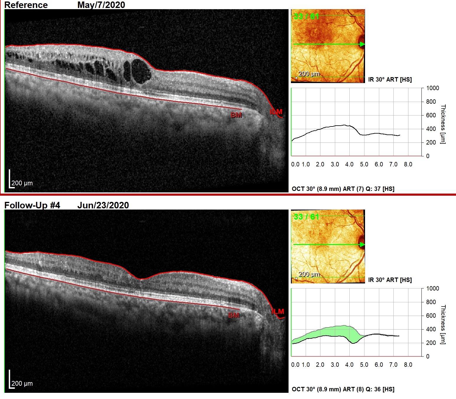
OCT, Optical coherence tomography maps of a patient who had a branch retinal vein occlusion, or BRVO.
The maps on the top show clinically significant macular edema causing a decrease in vision. The maps on the bottom show the same eye scanned a month later, utilizing state of the art eye tracking after anti-VEGF therapy. Substantially decreased leakage is present allowing for an increase in vision for this patient.
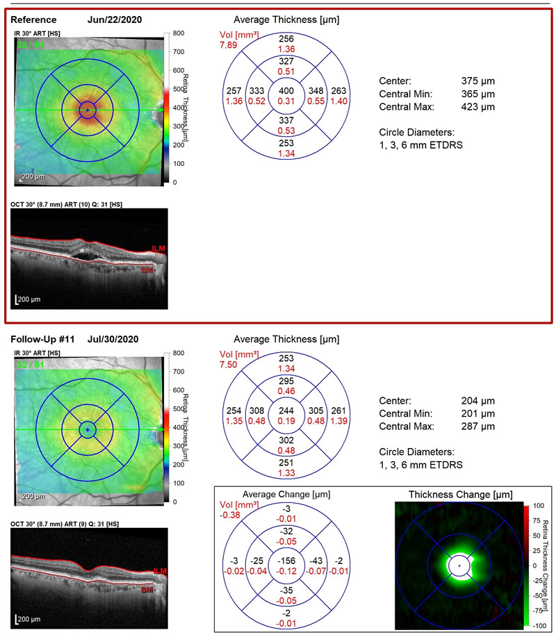
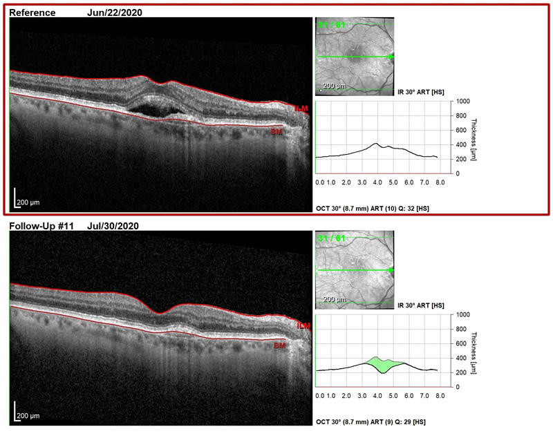
OCT, Optical coherence tomography maps of a patient who has wet macular degeneration, or wet AMD
The maps on the top are an example of a patient with wet macular degeneration. The maps show macular thickening with subretinal fluid involving the fovea (central vision) and the juxtafoveal retina consistent with wet macular degeneration.
The follow up thickness maps below show NO significant macular thickening, macular edema, or subretinal fluid after an ANTI-VEGF treatment. The comparison to retinal scans prior to treatment now show NO active leakage. Treatment was a success!


